Test Overview
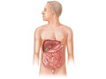
Magnetic resonance imaging (MRI) is a test done with a large machine that uses a magnetic field and pulses of radio wave energy to make pictures of organs and structures inside the belly. In many cases MRI gives information about structures in the body that cannot be seen as well with an X-ray, ultrasound, or CT scan.
For an MRI test, you are placed inside the magnet so that your belly is inside the strong magnetic field. MRI can find changes in the structure of organs or other tissues. It also can find tissue damage or disease, such as infection or a tumour. Pictures from an MRI scan are digital images that can be saved and stored on a computer for further study. The images also can be reviewed remotely, such as in a clinic or an operating room. Photographs or films of selected pictures can also be made.
In some cases, contrast material may be used during the MRI scan to show certain structures more clearly in the pictures. The contrast material may be used to check blood flow, find some types of tumours, and show areas of inflammation or infection.
Although MRI is a safe and valuable test for looking at structures and organs inside the body, it is more expensive than other imaging methods and may not be available in all medical centres.
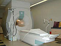
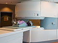
There are two main types of MRI—the standard MRI machine and the open MRI machine.
Why It Is Done
Magnetic resonance imaging (MRI) of the abdomen is done to:
- Find problems or tumours in the abdominal organs and tissues. In some cases, MRI can tell if a tumour is non-cancerous (benign) or cancerous (malignant).
- Check lower abdominal and pelvic organs for tumours, bleeding, or problems present since birth (congenital abnormalities).
- Find a blocked tube or stones in the tube that carry bile from the liver to the gallbladder (bile duct).
- Check organs and blood vessels prior to organ transplantation or surgery.
How To Prepare
For some MRI pictures of the belly, you may be asked to not eat or drink for several hours before the test.
Tell your doctor if you get nervous in tight spaces. You may get a medicine to help you relax. If you think you'll get this medicine, be sure to arrange a ride home. It may be unsafe for you to drive or get home on your own.
How It Is Done
Before the test
You will need to remove all metal objects (such as hearing aids, dentures, jewellery, watches, and hairpins) from your body. These objects may be attracted to the powerful magnet used for the test.
You will need to take off all or most of your clothes, depending on which area is examined. (You may be allowed to keep on your underwear if it's not in the way.) You will be given a gown to use during the test. If you are allowed to keep some of your clothes on, make sure your pockets are empty.
If you wear a medicine patch, you may need to remove it. The MRI can cause burns with some patches.
During the test
You will lie on a table that is part of the MRI scanner. Your head, chest, and arms may be held with straps to help you remain still. The table will slide into the space that contains the magnet. A device called a coil may be placed over or wrapped around the area to be scanned. A special belt strap may be used to sense your breathing. The belt triggers the machine to take the scan at the right time.
Some people feel nervous inside the MRI magnet. If feeling nervous keeps you from lying still, you can be given a medicine (sedative) to help you relax.
Inside the scanner, you will hear a fan and feel air moving. You may also hear tapping or snapping noises as the MRI scans are taken. You may be given earplugs or headphones with music to reduce the noise. It is very important to hold completely still while the scan is being done. You may be asked to hold your breath for short periods of time.
You may be given a medicine, such as glucagon, to slow bowel movements for some MRI tests.
During the test, you may be alone in the scanner room. But the technologist will watch you through a window, and you'll be able to talk back and forth.
If contrast material is needed, the technologist will put it in an IV in your arm or hand. The material may be given over 1 to 2 minutes. Then more MRI scans are done.
How long the
test takes
The test usually takes 30 to 60 minutes but can take as long as 2 hours.
How It Feels
You won't have pain from the magnetic field or radio waves used for the MRI test. The table you lie on may feel hard and the room may be cool. You may be tired or sore from lying in one position for a long time.
If a contrast material is used, you may feel some coolness when it is put into your IV.
In rare cases, you may feel:
- A tingling feeling in the mouth if you have metal dental fillings.
- Warmth in the area being examined. This is normal. Tell the technologist if you have nausea, vomiting, headache, dizziness, pain, burning, or breathing problems.
Risks
There are no known harmful effects from the strong magnetic field used for an MRI. But the magnet is very powerful. It may affect any metal implants or other medical devices you have.
Risks from contrast material
Contrast material that contains gadolinium may be used in this test. But for most people, the benefit of its use in this test outweighs the risk. Be sure to tell your doctor if you have kidney problems or are pregnant.
There is a slight chance of an allergic reaction if contrast material is used during the test. But most reactions are mild and can be treated using medicine.
If you breastfeed and are concerned about whether the contrast material used in this test is safe, talk to your doctor. Most experts believe that very little dye passes into breast milk and even less is passed on to the baby. But if you are concerned, you can stop breastfeeding for up to 24 hours after the test. During this time, you can give your baby breast milk that you stored before the test. Don't use the breast milk you pump in the 24 hours after the test. Throw it out.
Results
The radiologist may discuss initial results of the MRI with you right after the test. Complete results are usually available for your doctor in 1 to 2 days.
An MRI scan can sometimes find a problem in a tissue or an organ that is not seen by X-ray, ultrasound, or CT scan, even when the size and shape of the tissue or organ looks normal.
Magnetic resonance imaging (MRI) of the abdomen
|
Normal:
|
The organs and blood vessels are normal in size, shape, and location.
|
|
No abnormal growths, such as tumours, are present.
|
|
No blockage is found in the ducts draining the liver, gallbladder, or pancreas.
|
|
No blockage is found in the tubes (ureters) that lead out of the kidneys.
|
|
No bleeding, abnormal collections of fluid, blockage in the flow of blood, or bulges in the blood vessels (aneurysms) are present.
|
|
No signs of inflammation or infection are present.
|
|
Abnormal:
|
An organ is too large, too small, or in the wrong place. The MRI also may show areas of scarring or injury.
|
|
Growths are found, such as tumours that could be either benign or cancerous. Signs of infection may be present.
|
|
A collection of fluid is present, which could mean you have internal bleeding or an infection.
|
|
A bulge in the wall of a blood vessel (aneurysm) is present. Blockage in or narrowing of a blood vessel also may be found.
|
|
Blockage is present in the bile ducts. Reasons for the blockage may include a gallstone, tumour, infection, or inflammation.
|
|
Blockage is present in the tubes leading from the kidneys (ureters). Reasons for the blockage may include a kidney stone, tumour, infection, or inflammation.
|
Credits
Current as of:
December 19, 2022
Author: Healthwise Staff
Medical Review:
Kathleen Romito MD - Family Medicine
E. Gregory Thompson MD - Internal Medicine
Adam Husney MD - Family Medicine
Martin J. Gabica MD - Family Medicine
Howard Schaff MD - Diagnostic Radiology