An MRI is a test that uses a magnetic field and pulses of radio wave energy to make pictures of the spine. In many cases, an MRI gives different information than an X-ray, an ultrasound, or a CT scan. An MRI also may show problems that can't be seen with other imaging tests.
For an MRI, your body is placed inside a machine that contains a strong magnet. Pictures from an MRI can be saved and stored on a computer for further study. In some cases, a contrast material may be used during the MRI to show certain parts of the body more clearly.
The MRI can find changes in the spine and in other tissues. It also can find problems such as infection or a tumour. MRI can look at the spine in the neck (cervical), upper back (thoracic), or lower back (lumbosacral). The entire spine can be seen in one series of pictures to find a tumour. More detailed pictures of one area, such as the lumbar spine, may be taken.
Image courtesy of Intermountain Medical Imaging, Boise, Idaho. All rights reserved.
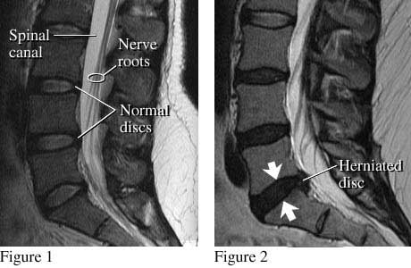
A side view of the lumbar spine shows normal discs, spinal canal, and nerve roots (see figure 1). Nerve roots normally float in the fluid-filled canal. Figure 2 shows a small herniated disc pushing into the canal toward nerve roots.
Health Tools
Health Tools help you make wise health decisions or take action to improve your health.
Decision Points focus on key medical care decisions that are important to many health problems.
Find problems of the spinal discs, such as a ruptured disc. The test may also show if a disc is pressing on a nerve, causing symptoms such as sciatica.
Find areas of the spine where the canal is abnormally narrowed (spinal stenosis) and may need surgery.
Find tumours affecting the bones or nerves of the spine. The tumours that most commonly spread to the spine include those from prostate, breast, or lung cancer.
Find compression fractures of the spine.
Check areas of joint inflammation (arthritis) or bone loss found during an X-ray test or a bone scan.
Find areas of the spine that do not have good blood supply.
Find an infection.
Find nerve damage caused by injury or disease, such as multiple sclerosis.
Check problems of the spine that have been present since birth (congenital).
An MRI may be done using contrast material to see abnormal tissue more clearly. The contrast material also may help tell the difference between old surgical scars and a new disease or injury.
In general, there's nothing you have to do before this test, unless your doctor tells you to.
Tell your doctor if you get nervous in tight spaces. You may get a medicine to help you relax. If you think you'll get this medicine, be sure you have someone to take you home.
How It Is Done
You may have contrast material (dye) put into your arm through a tube called an IV.
You will lie on a table that's part of the MRI scanner.
The table will slide into the space that contains the magnet.
Inside the scanner, you will hear a fan and feel air moving. You may hear tapping, thumping, or snapping noises. You may be given earplugs or headphones to reduce the noise.
You will be asked to hold still during the scan. You may be asked to hold your breath for short periods.
You may be alone in the scanning room. But a technologist will watch through a window and talk with you during the test.
How long the
test takes
The test usually takes 30 to 60 minutes but can take as long as 2 hours.
How It Feels
You won't have pain from the magnetic field or radio waves used for the MRI test. You may be tired or sore from lying in one position for a long time.
If a contrast material is used, you may feel some coolness when it is put into your IV.
In rare cases, you may feel:
Tingling in the mouth if you have metal dental fillings.
Warmth in the area being checked. This is normal. Tell the technologist if you have nausea, vomiting, a headache, dizziness, pain, burning, or breathing problems.
Risks
There are no known harmful effects from the strong magnetic field used for an MRI. But the magnet is very powerful. It may affect any metal implants or other medical devices you have.
Risks from contrast material
Contrast material that contains gadolinium may be used in this test. But for most people, the benefit of its use in this test outweighs the risk. Be sure to tell your doctor if you have kidney problems or are pregnant.
There is a slight chance of an allergic reaction if contrast material is used during the test. But most reactions are mild and can be treated using medicine.
If you breastfeed and are concerned about whether the contrast material used in this test is safe, talk to your doctor. Most experts believe that very little dye passes into breast milk and even less is passed on to the baby. But if you are concerned, you can stop breastfeeding for up to 24 hours after the test. During this time, you can give your baby breast milk that you stored before the test. Don't use the breast milk you pump in the 24 hours after the test. Throw it out.
Results
The radiologist may discuss some of the results of the MRI with you right after the test. Complete results are usually ready for your doctor in 1 to 2 days.
MRI of the spine
Normal:
The bones of the spine, discs, and nerves are normal.
No tumours, inflammation, or areas of nerve damage in the spine are present.
No disease or bone loss in the spine is present.
No ruptured discs are present. There are no structures pressing on a nerve.
No structural problems that have been present from birth (congenital problems) are found.
Abnormal:
Tumours, inflammation, or areas of nerve damage in the spine are present. A disease of the spinal cord, such as multiple sclerosis, is found.
Broken bones or bone loss in the spine caused by injury or disease, such as arthritis, is found.
One or more discs of the spine are bulging or ruptured or pressing on a nerve.
A condition that has been present from birth (congenital condition) is found in the spine or the vertebrae.
Credits
Current as of:
December 19, 2022
Author: Healthwise Staff Medical Review: William H. Blahd Jr. MD, FACEP - Emergency Medicine E. Gregory Thompson MD - Internal Medicine Adam Husney MD - Family Medicine Martin J. Gabica MD - Family Medicine Kathleen Romito MD - Family Medicine Howard Schaff MD - Diagnostic Radiology
Medical Review:William H. Blahd Jr. MD, FACEP - Emergency Medicine & E. Gregory Thompson MD - Internal Medicine & Adam Husney MD - Family Medicine & Martin J. Gabica MD - Family Medicine & Kathleen Romito MD - Family Medicine & Howard Schaff MD - Diagnostic Radiology
Call toll-free in B.C., 8-1-1, or 7-1-1 for the deaf and hard of hearing to get personalized assistance. Speak to a navigator who can guide you to reliable health information or connect you with a health professional. Translation services are available in over 130 languages.